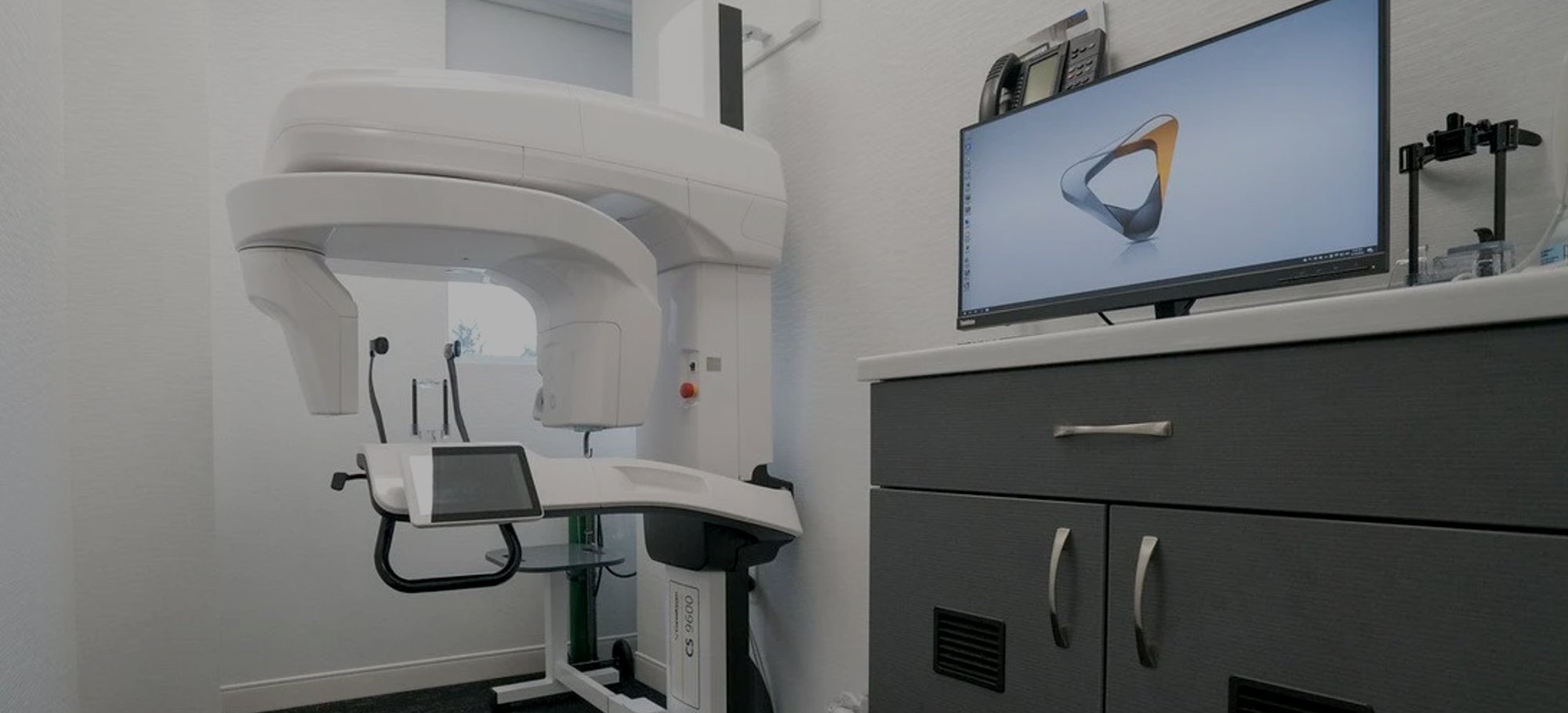
How to lower exposure to radiation in your practice
While it’s impossible to completely avoid radiation exposure in a dental practice, decreasing the risk for patients and staff should always be part of your dental radiography protocol.
Any radiation reduction methodology that can be practiced without major difficulty, expense or inconvenience, should be implemented.
Weighing the benefits
Maintaining a level of patient exposure that is as low as reasonably achievable begins with appropriate radiograph prescription. Contemplate diagnostic benefits in relation to increased exposure and carefully choose which X-rays will best allow for the detection of tooth and gum disease. For new patients, ask if a recent set of X-rays is on file with their old dentist prior to prescribing new radiography.
Rectangular collimation
Using an open-ended, shielded PID (position-indicating device) with rectangular collimation can reduce patient exposure by limiting beam size to the dimensions of the film. Round collimators can easily be converted to rectangular with an insert available from your preferred dental radiographic manufacturer.
Follow minimum coverage requirements
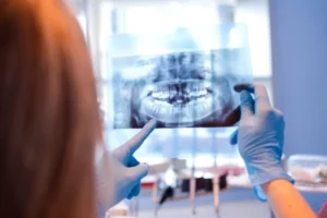
The area and volume of exposed tissue should always be limited to the bare minimum required for imaging the area in question. Standard guidelines exist for both periapical and bitewing techniques.
- Periapical: Include 1/4 inch of alveolar bone beyond the apex of each tooth and 1/8 to 1/4 inch margin between the crowns and the edge of the film.
- Bitewing: Strive for equal distribution of maxillary and mandibular crowns and alveolus. Make sure that interproximal spaces are open.
For both techniques, the occlusal plane ought to be straight or slightly curved in an upward slope toward the distal.
Converting from D-speed to F-speed film
Using X-ray equipment designed for short exposures allows for the use of group F film. F-Speed film can reduce radiation exposure up to 60% compared to group D. Further reduction can be achieved by closely following exposure guidelines for image density to adjust milliampere-seconds (mAs) accordingly.
Minimize errors in technique and processing
A common cause of unnecessary radiation exposure is human error in radiograph application. Faulty technique, improper adjustments or poor processing quality can all lead to retakes.
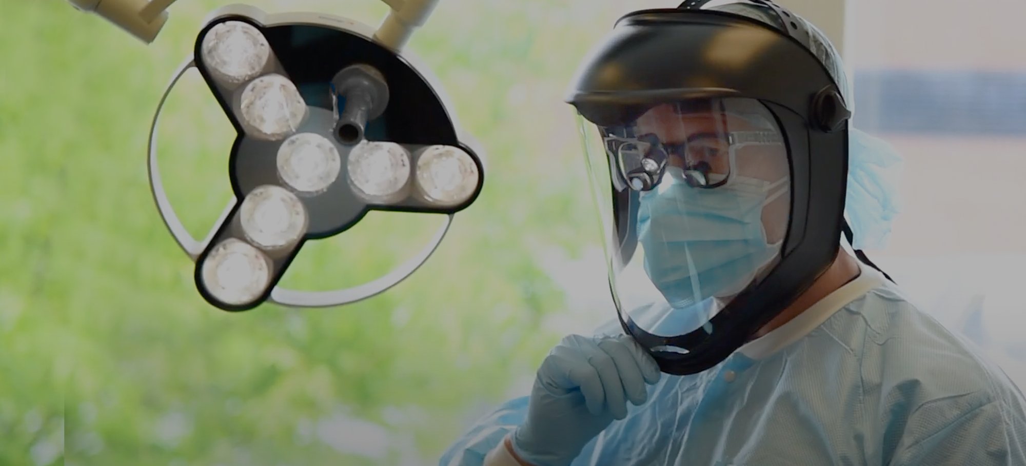
Exposure protection
Leaded lap aprons and thyroid collars should be used for all dental radiographs to guard against scattered radiation.
Protecting X-ray personnel is best achieved through the installation of a remote exposure button behind a radiation barrier (lead or concrete wall). In the event that a barrier is not available, the operator must stand a minimum of 6 feet from the X-ray tube head and at least 90 degrees from the primary beam.


